Mesothelioma imaging scans include X-rays, CT scans, MRIs and PET scans. They show doctors where tumors are, how far the cancer has spread and how well treatment is working. CT and PET are the most accurate for staging pleural mesothelioma. A biopsy is still required to confirm the diagnosis.
Diagnosed with mesothelioma? Get a free guide to learn about the latest treatment options.
Access information on top mesothelioma treatments.
Mesothelioma imaging scans help doctors spot tumors, pleural fluid and chest-wall thickening that point to cancer. The scan a specialist orders depends on where your symptoms are, your asbestos exposure history and what the care team needs to decide next.
Imaging Scans for Diagnosing Mesothelioma
CT scan: Shows detailed cross-section pictures to find tumors, map spread and guide biopsies.
MRI: Highlights soft tissues like chest wall, diaphragm and blood vessels to define local invasion.
PET scan: Lights up active cancer areas and helps stage disease and monitor response.
X-ray: A quick look for red flags like pleural effusions or thickening that warrant advanced imaging.
These tools work best together. Most patients start with a chest X-ray, then move to a contrast CT scan if results raise concern. These scans also help your doctor determine what stage the disease has progressed to and help develop a treatment plan based on that information. Imaging can find suspicious tumors, but only a tissue sample can confirm mesothelioma.
Expert Insight
“The first test for a patient with symptoms related to mesothelioma will be going to their primary doctor, getting an X-ray or a CT scan done. Based on the results from that, they may go through further testing such as a thoracentesis or paracentesis, which is where they drain fluid. The next step would be a biopsy.”
Dr. Snehal Smart, medical doctor and patient advocate at The Mesothelioma Center at Asbestos.com
Why Is Radiology Essential for Diagnosing Mesothelioma?
Mesothelioma radiology adds expert eyes to complex pictures. Radiologists trained in asbestos diseases can catch subtle signs, rule out look-alike problems such as pneumonia, and flag the safest spot for a biopsy. Their reports guide staging and treatment plans, and they partner closely with surgeons, medical oncologists and pulmonologists throughout care.
Imaging scans are also used for mesothelioma staging. Accurate staging requires determining the tumor’s size and location, whether lymph nodes are involved and if cancer cells have spread to other parts of the body.
Carla Fasolo first noticed mild back pain that quickly escalated to severe discomfort. An emergency X-ray revealed a large tumor in her chest, followed by a CT scan and biopsy that confirmed mesothelioma. Carla’s experience shows how imaging and pathology work together to detect mesothelioma early and guide treatment decisions.
Exclusive Content
Carla Fasolo: How did mesothelioma radiology diagnose your pleural mesothelioma?
I had pain in my back. And, It was not that uncomfortable, but it it was by my shoulder blade, and and it was annoying And I had my husband puts a medication on it, topical. And when he rubbed into it, I think he a little too hard because the pain became horrible.
And I ended up going to the doctor’s office, and they were gonna do an electrocardiogram. And he asked me to lay down, and I couldn’t lay down. The pain was unbearable.
So he told, his staff to call an ambulance, and my husband said it’s faster if taken by car. We went right to the hospital, the emergency room, they did an x-ray and came back and the doctor told me that I had a large tumor and that he didn’t know if it had metastasized or not. And so, I went to my doctor, back to my doctor, and he ordered a CAT scan.
Then they ended up doing a biopsy. And that’s how they found out that I had mesothelioma.
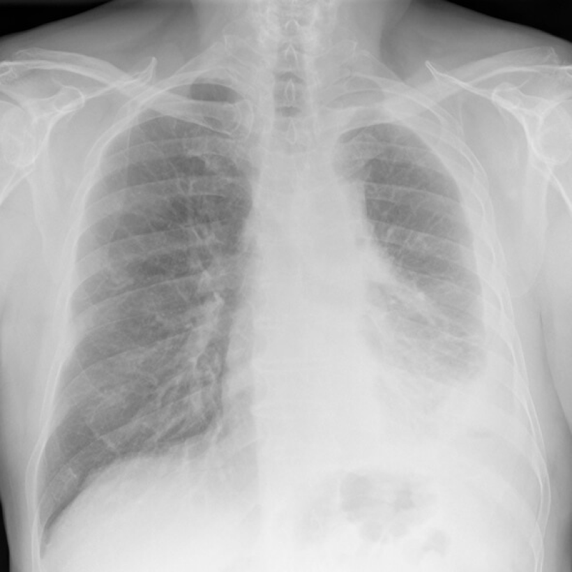
Mesothelioma X-Ray Scans
A chest X-ray is the first mesothelioma imaging scan most patients receive. It’s a fast, low-dose screen for pleural effusion, pleural thickening or masses that signal something is wrong. Doctors use them to spot fluid around the lungs, areas of unusual thickening or a collapsed lung that may signal pleural disease.
How X-Ray Scans Work
You stand or lie down in front of a flat detector and hold your breath for a few seconds.
The machine sends a brief, low-dose X-ray beam through your chest.
Bone blocks more of the beam, while air in the lungs lets it pass.
The detector catches this pattern and a computer turns it into a chest picture.
On the image, bone looks white, air looks black and fluid or thick tissue looks gray.
Doctors check for warning signs linked to mesothelioma, like pleural effusion, pleural thickening or a partly collapsed lung. X-rays can miss small or early tumors, and they do not diagnose cancer alone. If your X-ray raises concern, your doctor will order advanced scans and, if needed, a biopsy to be sure.
Michael Cole, a survivor of pleural mesothelioma, shares his X-ray story with us. A cough and lack of energy sent him to his doctor. Michael says, “They listened to my chest, took an X-ray and told me to go straight to the hospital. They quickly drained 2 liters of foul-looking fluid out of my right lung.” He was diagnosed with mesothelioma 4 days later.
What Does Mesothelioma Look Like on an X-Ray?
On a chest X-ray, pleural or pericardial mesothelioma tumors appear as wispy white areas around the lungs, while calcified tumors appear bright white. Bones appear white and healthy lungs are dark. Most abnormalities appear as lighter areas that are hazy or solid.
Tumors and scarring may distort chest anatomy. Compressed lungs or a raised diaphragm can be visible on an X-ray.
X-rays are 2D, making it hard to determine if a tumor is in the lung, pleura or the mediastinum around the heart. Additionally, X-rays don’t clearly show peritoneal or testicular mesothelioma. CT, MRI and PET/CT scans offer more detailed images for all mesothelioma types.
What X-rays can show include: fluid buildup, large masses and obvious pleural thickening. What X-rays can’t show include: small or early tumors and distinctions between mesothelioma and infections, scar tissue or benign asbestos-related pleural plaques. If symptoms persist, your doctor will order a CT scan. A normal X-ray doesn’t rule out mesothelioma and a biopsy is needed to confirm it.
500+ SPECIALISTS AVAILABLE
Get to the Right Mesothelioma Cancer Center, Faster
Connect with top-rated mesothelioma specialists at a cancer center near you, who will personalize treatment options based on your diagnosis.
A mesothelioma CT scan creates layered, cross-sectional images of the chest or abdomen that show tumors, lymph nodes and organs spread in detail. It’s the most-ordered imaging test doctors use to diagnose mesothelioma after an abnormal X-ray.
How CT Scans Work
You lie on a narrow table that slides through a short, donut-shaped scanner. The scanner sends thin X-ray beams around your chest from many angles.
Detectors measure the beams, and a computer builds “slice” pictures of your chest.
You may get contrast dye through an IV so the pleura, vessels and lymph nodes show clearly.
The technologist asks you to hold your breath for a few seconds to prevent blur.
They watch you from a window and talk through an intercom the whole time.
A CT scan is painless, takes only a few minutes and uses a small dose of radiation. A contrast CT can detect small tumors, measure pleural thickening, identify enlarged lymph nodes and check whether the cancer has spread to the diaphragm, chest wall or abdomen. Radiologists also use CT scans to plan biopsies and map out radiation treatment areas. CT scans sometimes miss how far a tumor has spread into nearby tissue, so doctors may add an MRI before surgery to get a clearer picture. If staging is still unclear, a PET/CT scan may be added.
Joey Barna is a pleural mesothelioma survivor. He spoke with us about his mesothelioma imaging scans. He shares, “I went to the hospital, and they thought I had pneumonia because they saw fluid on my lung. A week later I had a follow-up X-ray, and they saw that the fluid was still there. My primary doctor sent me for a CT scan. Fluid had built up and was pressing against my lung. It was preventing me from breathing properly.”
Expert Insight
“With the new staging system that we have for mesothelioma, it’s revolutionized things. We can actually measure on a CT and or PET scan how thick your tumor is. That can help guide a more aggressive or a less aggressive treatment.”
Dr. Jeffrey Velotta, thoracic surgeon at Kaiser Permanente Oakland Medical Center
PET Scans
Doctors inject a small amount of radioactive sugar into the bloodstream before a PET scan. Cancer cells absorb it faster than healthy cells, making tumors visible on the scan. When combined with a CT scan, a PET/CT is the most accurate way to stage pleural mesothelioma.
As Dr Jeffrey Velotta, a thoracic surgeon at Kaiser Permanente, tells us, “The PET scan is the most sensitive and specific test to tell you if you’ve had distant spread. We always get a CT and a PET scan.”
How PET Scans Work
A small amount of sugar-like tracer is put into your vein through an IV.
You rest quietly for about an hour while the tracer spreads through your body.
You lie on a table that moves through a ring-shaped scanner.
The scanner detects signals from the tracer where cells are very active.
A computer turns those signals into pictures that show “hot spots.”
For mesothelioma, PET/CT helps stage the disease, find hidden spread and measure how well treatment is working over time. A 2025 study in Neuroscience Informatics noted new patterns in PET/CT analysis that could soon improve the tracking of brain metastasis in mesothelioma.
Research suggests PET/CT outperforms CT and MRI alone for staging, giving doctors a clearer picture of tumor size, location and how far the disease has spread. That information shapes treatment planning and helps doctors gauge a patient’s outlook. Comparing scans over time also shows whether cancer is responding to treatment, holding steady or progressing, so doctors can adjust the plan as needed. PET/CT is expensive and not available everywhere, but it remains a key tool in mesothelioma care.
Karen R.
Verified Asbestos.com Survivor
Mesothelioma Survivor First Diagnosed Thanks to Optional Scan
During a regular annual checkup, Karen R. chose to have an optional heart scan even though her insurance didn’t cover it. It detected fluid around her lungs that led to a pleural mesothelioma diagnosis. She tells us, “If I had not chosen to get this optional scan the cancer would have gone undetected for much longer.”
A mesothelioma MRI uses powerful magnets, not radiation, to show soft tissues in unmatched detail, making it the preferred scan when surgeons need to know whether a tumor has invaded the chest wall, diaphragm or spine. A 2025 study in JTCVS Open noted it detected chest wall infiltration with more than 80% greater sensitivity than CT.
How MRI Scans Work
You lie on a table that slides into a short tunnel-shaped magnet.
The machine uses strong magnets and radio waves to make detailed pictures.
You must hold very still, and you may hear loud thumping sounds. Ear protection and a call button are provided in case you need help.
Sometimes an IV gives a contrast dye (gadolinium) to highlight tissues.
The technologist talks to you through an intercom the whole time. The scan is painless and usually takes 20 to 45 minutes.
MRI isn’t needed for everyone, but doctors may order one before surgery when the chest CT raises questions about chest-wall invasion, spinal involvement or diaphragm penetration, findings that change whether a tumor is resectable. MRI may also be used to re-evaluate after systemic therapy when CT findings are ambiguous. It isn’t used for routine surveillance because CT is faster and sufficient for most follow-up. Newer MRI techniques can detect smaller changes in tissue that older scans miss, giving doctors a more detailed picture of the disease.
Imaging Scans by Mesothelioma Type
Because different mesothelioma types start in different locations, not all patients benefit from all scans. There is no “best” mesothelioma imaging scan. Instead, doctors have an array of tools to fit pieces together at the right time to answer the right question.
CT and PET/CT are often used for whole-body imaging and staging. MRI and ultrasound are better for soft tissue and fluid details.
MRI
MRI scans can help locate testicular mesothelioma tumors earlier than X-ray or CT scans.
PET
PET scans can find very small tumors. This makes them very useful in pleural mesothelioma diagnosis and staging.
CT
CT scans can give doctors a more detailed look at pleural and peritoneal mesothelioma tumors.
X-Ray
X-ray scans can show abnormalities in the body, such as fluid buildup or masses.
Pleural Mesothelioma
For pleural mesothelioma, an X-ray is a quick first look at pleural effusion or pneumothorax. The primary test is a CT chest with contrast to observe pleural thickening, nodules and effusions. Ultrasound enables safe thoracentesis when assessing for fluid. Each of these scans flags signs of the disease and guides to the best site for biopsy.
To see tumors better, contrast CT highlights the pleural “rind” & lymphatics. PET/CT helps in staging, illuminating areas of active cancer and metastasis potential within and beyond the chest. An MRI is advantageous if the surgical team needs to know if a tumor invades the chest wall, diaphragm or spine.
Peritoneal Mesothelioma
For peritoneal mesothelioma, ultrasound can see ascites or fluid in the abdominal cavity and guide paracentesis. A CT of the abdomen/pelvis with contrast shows peritoneal thickening, omental caking and involvement with adjacent organs. These images guide treatment options and determine where biopsies should occur.
For visualization of tumors, a CT is still the gold standard since it assesses the abdomen rapidly, while an MRI, with specific diffusion sequences, can show smaller masses along the liver surface, within the spleen and in peritoneal folds. A PET-CT might help see disease outside the abdomen, though small serosal lesions may be more difficult to assess.
Pericardial and Testicular Mesothelioma
For pericardial mesothelioma, an echocardiogram is the quickest way to see if there is fluid around the heart and its influence on cardiac output. When pericardial thickening or a mass is suspected, either CT or MRI can confirm. Together, these findings best indicate if immediate drainage is needed and where, and rule out other causes of chest pain.
For visualization of pericardial tumors, cardiac MRI distinguishes the heart lining and reveals tumor extent. PET/CT helps stage outside the heart and into the lymphatics. For testicular mesothelioma, scrotal ultrasound is the first step to see if there’s a hydrocele or mass. Then, a CT and usually an MRI assess the spread to the abdominal or thoracic cavities.
Because pericardial and testicular mesothelioma are exceptionally rare, treatment planning at a specialized cancer center is critical. Most general oncologists won’t have direct experience with either subtype.
What to Expect During a Mesothelioma Scan
When you arrive for your mesothelioma imaging scan, the technologists will explain the process and answer your questions. Your doctor will likely recommend you wear loose, metal-free clothing or you’ll change into a gown. Bring a list of your medications and any allergies, especially to iodine or shellfish, which can react to CT contrast. Your doctor may also have you bring prior imaging on disc.
There are similarities in the process for each imaging type. You’ll need to lie flat on your back for most mesothelioma imaging. X-rays may involve standing or other positions. Staying very still is crucial for precise imaging. If you have limited mobility or pain, let the imaging team know.
Mesothelioma Imaging Scan Processes
Communication: CT and PET techs will talk to you via a speaker. For an MRI, headphones cancel noise and allow communication. Techs may tell you to hold your breath briefly.
Contrast dye: For CTs and MRIs you may be given a contrast dye orally or intravenously. This enhances image visibility.
Duration: A CT scan typically only takes about 10 minutes. PET scans and MRIs can take 30 to 90 minutes. Most patients can drive home after a CT or MRI, though some prefer to have someone pick them up after a PET scan because of the radioactive tracer.
Radioactive tracer: For PET scans, you’ll have an IV infusion of a radioactive tracer an hour before your scan.
Results: A radiologist reads most mesothelioma CT, PET and MRI results within 24 to 48 hours. Your mesothelioma doctor will discuss them and what they mean for your treatment plan at a follow-up appointment.
An MRI scan is similar to a CT, but it’s very loud. Metal coils in the scanner make booming and banging noises. People wear headphones throughout the scan. Also, the MRI scanner is more enclosed, which can cause anxiety. You will have a distress button to press if you need to stop the scan. If you have claustrophobia, ask your specialist about open MRI options or a mild sedative.
Before an MRI, tell your doctor and the technicians if you have any metal in your body. This includes surgical implants, pacemakers or shrapnel. An MRI’s strong magnetic field can affect metal objects. This could cause serious complications.
Exclusive Content
Tamron Little: How do you cope with scanxiety?
[MUSIC PLAYING] Scan anxiety is real. I get it every time. I can go for a scan of my toe, and I’m like, oh, my gosh. Because it’s just a traumatic experience and that you’re reliving it again. So when I do go for my scans, for my two-year scans or whenever I go, I do get scan anxiety. I do get itchy and sweaty. But once it’s over, everything’s clear. I’m good. But one of the things that I would suggest is to take a deep breath. Do not be so overly concerned. And don’t worry about anything that hasn’t happened yet. [MUSIC PLAYING]
Monitoring Mesothelioma Treatment With Imaging Scans
Mesothelioma patients typically have follow-up imaging every 3 to 6 months during the first year after treatment, then less often if the cancer remains stable. These surveillance scans show whether the cancer is shrinking, unchanged, growing or returning.
Survivor Tamron Little shares her scan experiences with us. She says, “The frequency of scans can depend on the patient and their progression. Before I had HIPEC surgery, I had a lot of preliminary scans.”
She adds, “After my surgery I had a follow-up CT scan a couple of weeks later. Then, after my doctor saw I was doing well with no cancer being found, it was 1 every 6 months. Then it was every 18 months up until the 5-year mark.”
Kevin Hession, a pleural survivor, says scans can pick up surgery’s effects. He tells us, “There is a lot of scar tissue around my left lung where they did a lot of work. Looking at it from the CT scans, you can see the scar tissue and it kind of surrounds the left lung.”
Common Questions About Mesothelioma Imaging Scans
Does asbestos show up on a CT scan?
No, asbestos fibers are microscopic. They aren’t visible on imaging scans. A high-powered microscope can see asbestos fibers in a biopsy. The presence of these fibers can help confirm a diagnosis of an asbestos-related disease.
Does mesothelioma show on a CT scan?
Yes. Mesothelioma typically shows on a CT scan as pleural thickening, pleural fluid (effusion), pleural plaques or a soft-tissue mass along the lining of the lung or abdomen. Contrast CT is the most commonly ordered imaging test for diagnosing and staging mesothelioma after an abnormal X-ray.
What is the best imaging scan for mesothelioma?
PET/CT is the most accurate scan for staging pleural mesothelioma because it shows both where tumors are and how active they are. CT is the standard first step for detection and follow-up. MRI is preferred when surgeons need to know whether a tumor has invaded the chest wall, diaphragm or spine. Most patients receive a combination of these scans.
What is the gold standard for diagnosing mesothelioma?
A surgical biopsy with a review from a pathologist who has mesothelioma expertise is the gold standard for diagnosing mesothelioma. Imaging scans like CT, PET and MRI find suspicious tumors, but only a biopsy can confirm the cancer cell type (epithelioid, sarcomatoid or biphasic), which determines treatment options and prognosis.
What are the risks or side effects of mesothelioma imaging scans?
X-rays use minimal radiation and are considered safe. CT scans involve significantly more, so repeat scans are avoided when possible. Contrast dye used in CT and MRI scans can cause allergic reactions or nausea in some patients. PET scans also involve radiation and a small risk of allergic reaction to the radioactive tracer. MRI can cause temporary effects from the magnetic field, including dizziness, nausea, a metallic taste or brief flashes of light. None of these scans are recommended during pregnancy.
Can ultrasound detect mesothelioma?
Ultrasound can be part of a mesothelioma diagnosis. But its use isn’t common. Soundwaves create its images. These soundwaves don’t travel well through air. So, it is hard to get good images in the chest cavity and abdomen.
While ultrasound’s images aren’t as detailed as CT scans or MRIs, they can be used to look at fluid buildup. Echocardiogram, for example, is a type of ultrasound. Doctors may use it if there’s fluid around the heart. This can help indicate an issue such as pericardial mesothelioma.
Recommended Reading
Your web browser is no longer supported by Microsoft. Update your browser for more security, speed and compatibility.
If you are looking for mesothelioma support, please contact our Patient Advocates at (855) 404-4592
Fact Checked
Our fact-checking process begins with a thorough review of all sources to ensure they are high quality. Then we cross-check the facts with original medical or scientific reports published by those sources, or we validate the facts with reputable news organizations, medical and scientific experts and other health experts. Each page includes all sources for full transparency.
Reviewed
Asbestos.com is the nation’s most trusted mesothelioma resource
The Mesothelioma Center at Asbestos.com has provided patients and their loved ones the most updated and reliable information on mesothelioma and asbestos exposure since 2006.
Our team of Patient Advocates includes a medical doctor, a registered nurse, health services administrators, veterans, VA-accredited Claims Agents, an oncology patient navigator and hospice care expert. Their combined expertise means we help any mesothelioma patient or loved one through every step of their cancer journey.
More than 30 contributors, including mesothelioma doctors, survivors, health care professionals and other experts, have peer-reviewed our website and written unique research-driven articles to ensure you get the highest-quality medical and health information.
About The Mesothelioma Center at Asbestos.com
Assisting mesothelioma patients and their loved ones since 2006.
Helps more than 50% of mesothelioma patients diagnosed annually in the U.S.
A+ rating from the Better Business Bureau.
5-star reviewed mesothelioma and support organization.
My family has only the highest compliment for the assistance and support that we received from The Mesothelioma Center. This is a staff of compassionate and knowledgeable individuals who respect what your family is experiencing and who go the extra mile to make an unfortunate diagnosis less stressful. Information and assistance were provided by The Mesothelioma Center at no cost to our family.
Asbestos.com. "Mesothelioma Imaging Scans." Last modified July 2, 2026. https://www.asbestos.com/treatment/scans/.
Most helpful, steered us in the right direction for treatment. Great source of information and support, the Center followed through on every one of our requests.
Because of their guidance, I was able to navigate getting my mother into MD Anderson when her case got put aside by mistake in all the COVID-19 craziness. Vanessa was amazing and I can’t recommend enough reaching out to them. I thought it was a gimmick to get you to hire a lawyer, but I was so wrong. They truly seemed to want to help meso patients and KNOW what you need to ask and do in order to get help.
Hearing the news about my mother's diagnosis was heartbreaking. I felt lost, I didn't know how I could help or where to seek the best medical care. I started researching specialists online and shortly after, Dr. Smart reached out to me. She has been extremely helpful and encouraging throughout this entire process. Even though we aren't located in her area, she has helped us get in contact and set up appointments with the best doctors/specialists nearby. She has always been available for any questions that we have, and she even sent us a binder full of helpful resources. The patient advocates are amazing and true to their title. Dealing with this process is not easy, but knowing that we have someone like Dr. Smart in our corner is reassuring and we are so grateful for her and The Mesothelioma Center.
My son Carlos was diagnosed with this terrible and unknown disease a few months ago. Thank God we found The Mesothelioma Center along the way, and Vanessa Blanco who provided us with information on hospitals and doctors who have been of great help. I am very grateful to them.
Extremely communicative and helped my dad get an appointment with one of the top centers in Philadelphia. I'm so grateful for this center. They assisted with information on nutrition, legal help, and scheduling appointments. Special thanks to Danielle!
Danielle DiPietro was an invaluable resource for me. Her suggestions and recommendations guided us towards stellar practitioners in our area. Without her advocacy, I feel we would have been receiving less-than-optimal medical and legal care for mesothelioma. Receiving the diagnosis was a shock and I felt lost initially. I wish everyone could take advantage of this FREE assistance.
I was very grateful and appreciative of Dr. Smart from The Mesothelioma Center. She was very helpful to my husband and me. She educated and walked us through the steps, and suggested ideas and questions to ask his doctors. She also provided me with a lot of information that I can read and educate myself about this illness. We need more people like Dr. Smart, who is very educated and you can tell she enjoys the work that she does by the way she assisted my husband and me. We thank the Lord and are grateful that we met Dr. Smart from The Mesothelioma Center.
In January of 2016, my husband was diagnosed with peritoneal mesothelioma. Our first reaction was: what is this and what can we do? He was diagnosed by an oncologist and was scheduled to start chemotherapy. When we arrived home that day, I googled mesothelioma and discovered The Mesothelioma Center had a form to fill out to request additional information. I filled it out and within an hour, I received a phone call from Karen Selby from The Mesothelioma Center asking if I needed any help. Karen was and still is my lifeline. She located a doctor at the Cleveland Clinic who performed surgery and HIPEC on peritoneal mesothelioma patients. My husband was scheduled with an appointment and his surgery was performed on March 3, 2016. He continued with follow-up appointments with the oncologist until a friend of ours passed away from it in 2017. Immediately I sent Karen an email asking if she knew any mesothelioma specialists at the clinic, and of course, I got a prompt response back with a name. Everything was going well until the last CAT scan, which showed it returned. He is now doing chemo and has his next CAT scan scheduled for the end of March with a follow-up for the results with the mesothelioma doctor. Without Karen, I am not sure my husband would still be here. She provided me with so much information along with help in various ways, too numerous to even mention. Thank you to all those who are there to help us.
Dr. Landau is the Medical Director of Virtual Hematology at the Medical University of South Carolina, where he leads programs that expand access to cancer care through telehealth. With more than 18 years of experience in oncology and hematology, he specializes in hematologic and genitourinary cancers, including bladder, prostate and kidney cancers. He has held multiple leadership roles in cancer program development and previously served as section chief of hematology and oncology at Orlando Health UF Health Cancer Center, where he founded its telehealth program.
Fact-checked and verified content:
Our fact-checking process begins with a thorough review of all sources to ensure they are high quality. Then we cross-check the facts with original medical or scientific reports published by those sources, or we validate the facts with reputable news organizations, medical and scientific experts and other health experts. Each page includes all sources for full transparency.
Please read our editorial guidelines to learn more about our content creation and review process.