Navy veteran Jerry Cochran was diagnosed 50 years ago with sarcoidosis. The more he read about it, the more concerned he was that his symptoms just didn’t match up. In 1991, he was given a new diagnosis – silicosis – from inhaling silica dust while scraping paint on the USS Independence. He was also later diagnosed with asbestosis, an asbestos-related interstitial lung disease.
Interstitial Lung Disease & Asbestos
Interstitial lung disease causes inflammation and scarring. Asbestos exposure, some diseases and medications can cause it. There is a clear link between asbestos exposure and asbestosis or interstitial pneumonitis, a form of ILD.

What Is Interstitial Lung Disease?
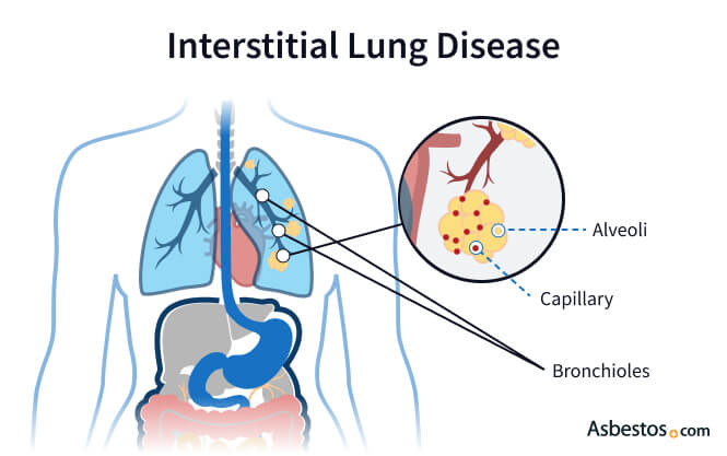
Interstitial lung disease is an umbrella term for approximately 200 relatively rare lung diseases that cause lung inflammation or scarring. Some ILD types only cause scarring (fibrosis). Some only cause inflammation, and others cause both.
Key Facts About Interstitial Lung Disease
- Genetics, asbestos exposure, lifestyle factors, medications and other health conditions can cause interstitial lung disease.
- ILD covers diseases that scar the lungs.
- Symptoms may take years to develop. They can cause coughing, fatigue and shortness of breath.
- You can’t reverse lung damage, but early treatment can slow or stop its progression.
If a patient has inflammation without scarring, early treatment may prevent scarring. The inflammation and scarring affect the interstitial lining that supports the lung’s alveoli. This decreases lung function as thickened tissue can’t supply oxygen to the blood as effectively as healthy lung tissue.
Asbestosis is an ILD linked to asbestos exposure. As the body tries to expel inhaled asbestos fibers, inflammation and scarring occurs. It’s a progressive disease that continues to worsen over time.
Types of Interstitial Lung Disease
ILDs are usually grouped by their cause into broad categories. These categories include exposure to irritants, some types of medical treatment, autoimmune disorders and smoking.
The causes of some ILD cases can’t easily be identified. They are often called idiopathic pulmonary fibrosis. Idiopathic means the disease was spontaneous or the cause is unknown.
Types of ILDs
- Desquamative interstitial pneumonitis: Cigarette smoking can cause many serious, fatal illnesses. It is responsible for this form of ILD.
- Hypersensitivity pneumonitis: This type of ILD comes from inhaling irritants like dust or mold.
- Idiopathic pulmonary fibrosis: This is a chronic and progressive fibrosis from an unknown cause.
- Interstitial pneumonia: Bacteria, fungi or virus exposure can cause an often temporary condition with inflammation of the interstitium.
- Sarcoidosis: An inflammatory disease that mainly affects the lungs and lymph nodes. It can also affect other organs.
Autoimmune disorders contributing to ILD include lupus, polymyositis or dermatomyositis, rheumatoid arthritis and scleroderma. Risks include exposure to asbestos, silica dust and cotton dust. Some medical treatments may cause lung inflammation and scarring. These treatments include chemotherapy, radiation and certain medicines.

Understand your diagnosis, top doctors and ways to afford care.
Get Your Free GuideInterstitial Lung Disease Symptoms
Many of the different forms of ILD display similar symptoms associated with decreased lung function. They usually appear after scarring develops. This means that irreversible damage has occurred.
Symptoms of asbestosis can easily be mistaken for signs of other lung conditions such as COPD or asthma. Many ILD symptoms worsen with exertion.
Interstitial Lung Disease Symptoms
- Chest pain or discomfort
- Clubbed fingers
- Difficulty or labored breathing
- Dry cough
- Fatigue and weakness
- Fever
- Shortness of breath
- Unexplained loss of appetite or weight
Latency periods vary among the types of interstitial lung disease. Symptoms can take months to years to appear, depending on the disease. Some ILDs have a long latency period, so symptoms may not appear right away. Others advance more rapidly.
It can take 20 to 30 years for asbestosis symptoms to develop. Some patients have no symptoms at all. Many patients don’t know they have the condition until lung damage becomes significant many years after exposure.
Complications
Patients with ILD can suffer from serious complications. Many ILD complications result from decreased lung function and its impact on the body. They include fluid and calcium buildup in the lung lining, high blood pressure in the lungs and a higher risk of infection and some cancers.
Interstitial Lung Disease Complications
- Collapsed lungs
- Lung infections
- Pleural effusions
- Pleural plaques
- Pulmonary hypertension
- Venous thromboembolism
Lung cancer is a big concern for asbestosis patients who smoke. Quitting smoking can lower your cancer risk and boost lung function.
Additionally, acute exacerbation is a severe concern. This sudden worsening of respiratory symptoms affects a large number of patients with ILD and can occur at any time. For many patients, it can be what ultimately leads to a diagnosis.
Interstitial Lung Disease Diagnosis
Doctors use chest X-rays, CT scans and lung tests to diagnose interstitial lung diseases. The symptoms of ILDs and other lung conditions are similar. This can make diagnosis difficult. Comprehensive testing can help rule out other conditions, so doctors can make a proper diagnosis.
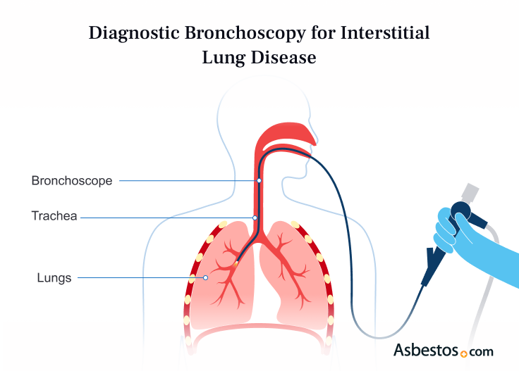
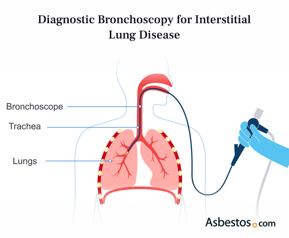
In some cases, a lung biopsy may be necessary. Tissue collected can show the condition of the lungs. It may also detect other lung conditions like cancer. Doctors collect samples using a bronchoscopy, lavage or biopsy.


Treatment for Interstitial Lung Diseases
ILD and asbestosis treatment aims to relieve symptoms and complications. It also slows lung scarring. Oxygen can help with shortness of breath from damage. Other medications may help. These include corticosteroids, anti-fibrotics and tyrosine kinase inhibitors. They may help with some symptoms and slow the disease.
More intensive interventions include draining fluid from the lungs and respiratory therapy. In the latter, a trained therapist teaches patients breathing exercises to improve lung function. In rare cases, a lung transplant may be needed. But it isn’t a common treatment for asbestosis.
ILD & Asbestosis Treatment Options
- Lifestyle changes
- Oxygen therapy
- Prescription medications
- Pulmonary rehabilitation
Prognosis after an ILD diagnosis varies greatly by disease type. General physical condition and certain lifestyle factors, such as smoking, can impact disease progression. Smokers exposed to asbestos are very likely to develop ILD.
With asbestosis, a patient’s prognosis often depends on the length and severity of asbestos exposure. Individuals may live full lives for many years after a diagnosis.
Recommended Reading


