Epithelioid mesothelioma is the most common subtype of malignant mesothelioma, accounting for 50% to 70% of all cases. Caused by asbestos exposure, it has the best prognosis and most treatment options of all mesothelioma cell types.
Learn more about mesothelioma, asbestos and the steps you can take to fight this disease.
About 54% of pleural mesothelioma tumors contain epithelial cells.
Doctors diagnose 1,500 to 2,100 patients with epithelioid mesothelioma annually.
Epithelioid cells comprise about 75% of peritoneal mesothelioma tumors.
The epithelioid type has the best prognosis of all mesothelioma subtypes.
Average life expectancy with surgery is approximately 18 months.
The latency period is 20 to 60 years after initial asbestos exposure.
What Is Epithelial Mesothelioma?
Epithelial mesothelioma is an aggressive form of mesothelioma cancer that develops on the tissue that lines the organs of your body. This membrane, called the mesothelium, is made of cells like epithelial cells. Inhaled asbestos fibers can get stuck in that membrane and cause irritation that changes the DNA of the cells, making them cancerous.
Pathologists look at tumor and tissue samples and determine what cells they can see. If the majority of cells from your mesothelioma sample are epithelioid cells, your mesothelioma subtype is determined to be epithelioid mesothelioma.
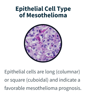
“Epithelioid” and “epithelial” are sometimes used in ways that feel interchangeable. But epithelial cells are normal cells that line the skin and the inside of organs. Epithelioid cells can be found in both cancerous and non-cancerous conditions. Epithelioid cells look like epithelial cells, which are shaped like columns or cubes. However, epithelioid cells may come from different types of cells that have mutated or changed shape, taking on an appearance similar to epithelial cells.
The epithelioid subtype of mesothelioma has a more favorable prognosis. Epithelioid cells respond better to aggressive treatment than sarcomatoid cells. Having a combination of epithelioid and sarcomatoid cells is called biphasic mesothelioma. People with this subtype benefit if they have more epithelioid than sarcomatoid cells.
Epithelioid Mesothelioma Symptoms
Epithelioid mesothelioma symptoms often include cough, shortness of breath and lack of appetite. Symptoms can depend on where on the mesothelium the cancer develops: the lining of the lungs (pleura), abdomen (peritoneum), heart (pericardium) or testes (tunica vaginalis).
Common Symptoms of Epithelioid Mesothelioma
Abdominal pain or bloating
Ascites or fluid buildup in the belly (peritoneal effusion)
Bowel or bladder changes
Chest tightness or pain
Cough, hoarseness or difficulty swallowing
Difficulty breathing
Fatigue
Fever or night sweats
Fluid buildup in the chest (pleural effusion)
Loss of appetite
Nausea, vomiting or diarrhea
Shortness of breath
Unexplained weight loss
Studies show early symptoms may be mild, making it difficult to diagnose. Late diagnosis can cause delays in treatment. As the disease progresses and tumors increase in size and spread, more severe symptoms may appear.
Mesothelioma cancer symptoms are the same, no matter the cell type. But the cell type affects which treatments are most helpful. Talk to your doctor if you experience any of these symptoms.
500+ SPECIALISTS AVAILABLE
Get to the Right Mesothelioma Cancer Center, Faster
Connect with top-rated mesothelioma specialists at a cancer center near you, who will personalize treatment options based on your diagnosis.
The primary risk factor and cause of epithelioid mesothelioma is asbestos exposure. This is true for other types of mesothelioma as well. Inhaling or swallowing asbestos fibers causes inflammation and DNA damage. This can lead to cancer many years later.
People are commonly exposed to asbestos at work, in the environment, from contaminated talc or damaged asbestos products in homes, schools or other older buildings. Not everyone exposed to asbestos develops mesothelioma, though there is no safe amount of exposure. Additional risk factors can include the amount and length of exposure and possible genetic predispositions for developing cancer.
Many people with epithelioid mesothelioma worked with asbestos products years before their diagnosis. All mesothelioma cell types have a latency period of 20 to 60 years. The first symptoms may not appear for decades after the initial asbestos exposure.
Christine S.
Verified Asbestos.com Survivor
Survivor Faces Malignant Epithelial Mesothelioma
Christine S. was diagnosed with epithelial mesothelioma after nearly a year of unanswered questions and pleurisy and pneumonia misdiagnoses. She remained resilient through 6 intense rounds of chemo. Today, she shares her experience to raise awareness about the risks of secondary asbestos exposure and advocate for earlier diagnosis of mesothelioma.
A tissue biopsy is the only way to diagnose epithelioid mesothelioma. This procedure involves taking samples of suspicious tissue. Pathologists examine the tissue samples under a microscope to identify specific cell characteristics.
Epithelioid mesothelioma cells clump together in groups and don’t tend to travel. These cell types are less likely to spread to other areas of the body. When a pathologist confirms the presence of specific cancer cells, an accurate diagnosis of your mesothelioma type can be made.
Your mesothelioma doctor will review your detailed personal pathology report that describes the types of mesothelioma cells found. You and your doctor will discuss the types of treatments that typically work best for epithelioid mesothelioma, your overall health and goals to determine which therapy options may be best for you.
Exclusive Content
Insights: How to Test for Mesothelioma
We actually have this amazing brochure that we can send to a patient that thinks they might have mesothelioma, never been to a doctor before, never been diagnosed. We offer a brochure they literally can take to the doctor’s office with them and literally hand it to the doctor and say, I think I have this. Can I have these tests?
The first test for a patient with, symptoms related to mesothelioma will be going to their primary doctor, getting an x-ray or a CAT scan done. Based on the results from that, they may go through further testing such as a thoracentesis or paracentesis, which is where they drain fluid, whether it’s from the lining of the lungs or the peritoneal area, which is the abdominal area. The next step would be for a biopsy. The biopsy is a step where they confirm the diagnosis. This sample is sent to a pathologist who ultimately tests the sample taken out from the tumor and identifies it as mesothelioma.
And not just look at under microscope by any pathologist, but pathologist who has an expertise in making that diagnosis. Because even with a good sample of tumor, looking at it under a microscope, it can sometimes be difficult to make that diagnosis. And it’s important not only to know that it’s mesothelioma, it’s important to know what is the histological subtype. Is it epithelial or is it sarcomatoid or is it a mixture of both called mixed or biphasic, because that changes the type of treatment.
We handhold them through the diagnostic part early on. So we actually prepare them before they go of what their doctor is likely going to do or what may not need to do based on their previous studies they’ve had.
Diagnosing Epithelial Mesothelioma With Immunohistochemistry
The tool or technique for studying cancer tissues is called immunohistochemistry. Pathologists look at stained samples, testing for certain proteins linked to epithelial cells. If pathologists find proteins from other cancers, they’ll rule out epithelioid mesothelioma.
The proteins that help doctors identify epithelioid mesothelioma from different types of cancer include: calretinin, D2-40, keratin 5/6, podoplanin and WT-1 protein. An official diagnosis depends on more than just immunohistochemistry. It also considers the tumor’s appearance, location and cell traits.
Epithelial subtype mesothelioma describes the type of cells the pathologist is seeing under the microscope when they look at a patient’s tumor.
Dr. Andrea Wolf, director of the New York Mesothelioma Program at Mount Sinai
Subtypes of Epithelial Mesothelioma Cells
While epithelioid is a subtype of mesothelioma, there are further subtypes of the epithelioid type. Pathologists can identify these cell subtypes with immunohistochemistry.
Epithelioid mesothelioma has a better prognosis than other subtypes, but some epithelioid cell subtypes also have better prognoses than others. For example, adenomatoid cells are associated with a better mesothelioma survival rate.
Patient Advocate Danielle DiPietro explains why it’s so important to diagnose your cell subtypes. Daniells tells us, “When I speak with a patient who is hesitant to have a biopsy, which is rare, I explain that diagnosing the cell type is imperative for assessing treatment options.”
Adenomatoid
Also known as the microglandular cell type, this accounts for only 6% of pleural cases. The peritoneal form behaves like benign lesions and responds well to treatment.
Deciduoid
Doctors have diagnosed fewer than 50 cases of this very rare subtype. It most often affects young women. Just more than 50% of deciduoid cases occur in the abdomen, and less than 50% occur in the pleura. It can be mistaken for other cancers, including a type of lung cancer known as squamous cell.
Glandular
Glandular tumors often develop in the pleura and have patterns that resemble glands. These cells behave like a type of cancer called adenocarcinoma that has spread to the pleura.
Small Cell
Small cell mesothelioma doesn’t show the patterns found in small cell lung cancer. Those patterns include stream, ribbon or rosette. This cell type occurs with greater frequency in the abdomen. The survival rate is around 8 months.
Solid
Well-differentiated solid cells group in nests, cords or sheets. They resemble noncancerous abnormal cell growth. Poorly differentiated cells may look like large cell carcinoma or lymphoma.
Tubulopapillary
This common epithelial subtype can resemble a cancer called adenocarcinoma of the pleura. It’s not the same as benign well-differentiated papillary mesothelioma.
Well-Differentiated Papillary Mesothelioma
WDPM is a rare subtype of mesothelioma often found in young women with no history of asbestos exposure. It’s also less likely to spread or cause significant harm compared to other types of mesothelioma.
How Is Epithelioid Mesothelioma Treated?
Doctors often use a mix of treatment options like chemotherapy, immunotherapy and surgery to treat epithelioid mesothelioma. Using different therapies together can help patients live longer.
People diagnosed in early stages usually undergo aggressive treatment, while those with late-stage cancer often opt for palliative care, which may include immunotherapy, chemo and Tumor Treating Fields. However, palliative care can be helpful at any stage to manage symptoms and improve quality of life.
Common Epithelioid Mesothelioma Treatments
Chemo: Chemo drugs penetrate and attack mesothelioma cells, either destroying them or preventing their spread. This treatment can triple the mesothelioma survival rate.
Combinationtherapy: Mixing treatment approaches, or multimodal therapy, integrates multiple mesothelioma treatments to fight cancer from different angles.
Radiation: Ionizing radiation, including high-energy X-rays or particles, is used to destroy cancer cells. This helps shrink tumors, relieve pain and reduce the risk of recurrence and spread.
Surgery: Procedures like extrapleural pneumonectomy or EPP and pleurectomy with decortication P/D are common surgeries for pleural mesothelioma. Tumor-removing surgery such as cytoreduction with hyperthermic intraperitoneal chemotherapy, known as HIPEC, is used for peritoneal mesothelioma.
The epithelioid cell type responds better to surgery and chemo than sarcomatoid or biphasic mesothelioma, affording mesothelioma patients more multimodal therapy options. As Dr. Jacques Fontaine, thoracic surgeon and director of the Mesothelioma Treatment and Research Institute at Moffitt Cancer Center, tells us: “Usually epithelial type is a slightly less aggressive type of tumor. And usually it responds fairly well to chemotherapy. It may even respond to immune therapy. Surgery may be an option in epithelial type mesothelioma in specific circumstances.”
Patient Advocates can connect you to experienced mesothelioma specialists at top cancer centers who can provide additional insights or offer different treatment choices. Patient Advocates or your healthcare team can also help you find clinical trials that test new therapies or improvements to standard treatments.
FREE MESOTHELIOMA GUIDE
Expert Guidance for Your Mesothelioma Journey
Get answers about treatment, top doctors, and financial support from the nation’s most trusted mesothelioma resource.
Epithelial Mesothelioma Life Expectancy and Prognosis
The average life expectancy for people with epithelioid mesothelioma who undergo surgery is about 18 months. Where the cancer develops affects life expectancy. People with pleural epithelioid mesothelioma who undergo surgery live an average of 18 months, while those with peritoneal disease have a median survival of nearly 5 years. Across both forms, survival can stretch from roughly 1.5 to 6.5 years depending on tumor location, stage at diagnosis and treatment response.
Epithelioid Mesothelioma Survival Rate
Overall 5-year survival rate: 12% for pleural epithelioid mesothelioma, which is higher than biphasic or sarcomatoid subtypes (Source: National Cancer Institute)
Survival with surgery: Up to 15% at 5 years for patients with resectable epithelioid MPM (Source: Cancer Medicine, National Cancer Database analysis)
2-year survival with chemo: ~35% among epithelioid patients, compared to lower rates in other subtypes (Source: Cancer Medicine)
Median survival with Imfinzi + chemo: 24.3 months for epithelioid patients, compared to 9.2 months for nonepithelioid, with a 65.9% response rate (Source: NCI)
Another study on peritoneal mesothelioma found that people with epithelioid cells lived a median of about 5 years. On average, people with epithelioid mesothelioma live 200 days longer than those diagnosed with other cell types.
Overall, epithelioid mesothelioma has a more favorable prognosis than biphasic or sarcomatoid mesothelioma. Epithelioid cells typically respond best to treatment and tend to spread more slowly, which contributes to longer survival.
Expert Insight
The survival rate for epithelial patients is overall significantly longer. Epithelial cell type is the best mesothelioma cell type to have. This patient will have more options, such as surgery, and is more likely to respond to treatment.
Karen Selby, Patient Advocate and registered nurse
Common Questions About Epithelioid Mesothelioma
Where can I get treatment for epithelial cell mesothelioma?
Look for mesothelioma specialists with years of experience treating peritoneal and pleural mesothelioma. Oncologists need training to treat epithelioid mesothelioma, and our Patient Advocates can help you find a top doctor.
Is epithelioid mesothelioma curable?
Epithelioid mesothelioma has no cure, but patients with this cell subtype have the most treatment options and respond best to all forms of treatment.
Is there any ongoing research on epithelioid mesothelioma?
Researchers are constantly looking for mesothelioma patients with the epithelioid cell type for clinical trials. As of May 2026, clinical trials are actively recruiting mesothelioma patients across the U.S. Studying epithelioid patients lets researchers learn how a drug or therapy affects most people diagnosed with mesothelioma. For example, a clinical trial completed in 2024 examined the effectiveness of Itraconazole and Rifampin on epithelioid mesothelioma patients. Final results haven’t been published.
How is epithelioid mesothelioma different from sarcomatoid mesothelioma?
Epithelioid mesothelioma differs from sarcomatoid mesothelioma in cell shape, growth pattern and treatment response. Epithelioid cells cluster together and spread more slowly, giving patients more treatment options and a more favorable prognosis. Sarcomatoid cells have a spindle shape, grow more aggressively and respond less well to chemotherapy. Doctors call a mix of both biphasic mesothelioma.
What are the subtypes of epithelioid mesothelioma?
Epithelioid mesothelioma includes 7 recognized subtypes: adenomatoid, deciduoid, glandular, small cell, solid, tubulopapillary and well-differentiated papillary mesothelioma. Each subtype has a distinct cell appearance under a microscope. Some subtypes, like adenomatoid, have a more favorable survival rate. Well-differentiated papillary mesothelioma is rare and typically less aggressive.
Recommended Reading
Your web browser is no longer supported by Microsoft. Update your browser for more security, speed and compatibility.
If you are looking for mesothelioma support, please contact our Patient Advocates at (855) 404-4592
Fact Checked
Our fact-checking process begins with a thorough review of all sources to ensure they are high quality. Then we cross-check the facts with original medical or scientific reports published by those sources, or we validate the facts with reputable news organizations, medical and scientific experts and other health experts. Each page includes all sources for full transparency.
Reviewed
Asbestos.com is the nation’s most trusted mesothelioma resource
The Mesothelioma Center at Asbestos.com has provided patients and their loved ones the most updated and reliable information on mesothelioma and asbestos exposure since 2006.
Our team of Patient Advocates includes a medical doctor, a registered nurse, health services administrators, veterans, VA-accredited Claims Agents, an oncology patient navigator and hospice care expert. Their combined expertise means we help any mesothelioma patient or loved one through every step of their cancer journey.
More than 30 contributors, including mesothelioma doctors, survivors, health care professionals and other experts, have peer-reviewed our website and written unique research-driven articles to ensure you get the highest-quality medical and health information.
About The Mesothelioma Center at Asbestos.com
Assisting mesothelioma patients and their loved ones since 2006.
Helps more than 50% of mesothelioma patients diagnosed annually in the U.S.
A+ rating from the Better Business Bureau.
5-star reviewed mesothelioma and support organization.
My family has only the highest compliment for the assistance and support that we received from The Mesothelioma Center. This is a staff of compassionate and knowledgeable individuals who respect what your family is experiencing and who go the extra mile to make an unfortunate diagnosis less stressful. Information and assistance were provided by The Mesothelioma Center at no cost to our family.
Asbestos.com. (2026, May 7). Epithelioid Mesothelioma. Retrieved May 28, 2026, from https://www.asbestos.com/mesothelioma/malignant/epithelial/
MLA
"Epithelioid Mesothelioma." Asbestos.com, 7 May 2026, https://www.asbestos.com/mesothelioma/malignant/epithelial/.
Chicago
Asbestos.com. "Epithelioid Mesothelioma." Last modified May 7, 2026. https://www.asbestos.com/mesothelioma/malignant/epithelial/.
The Patient Advocates are absolutely amazing and helpful! I am so very pleased with the information and the emotional support that I have received from everyone! When I felt all hope was lost, Amy gave me great information and was so compassionate. I do not feel alone, and I know there is strong support for anyone dealing with this tragic disease. I received a FREE information package, including a recipe book, information on the disease, a toll-free number to contact for assistance, and a book with the answers to 100 questions that are commonly asked. This information alone was worth my time! I read for hours and hours, and just that alone—knowledge—can be a great comfort. To fully understand this disease, to just have the knowledge!!! Thank you all!! Thank you so, so much.
I found out about The Mesothelioma Center while researching peritoneal mesothelioma days after being diagnosed with the malignant cancer. While a lot of the 'Mesothelioma' websites are actually law firms just trying to get your business, theirs is a user-friendly, comprehensive website set up to aid patients, their loved ones, and/or caregivers with all things related to a cancer diagnosis from asbestos exposure. I was soon put in touch with one of their doctors, Snehal Smart, M.D., who immediately sent me a packet of general information, a book on surviving mesothelioma, and offered me assistance in finding doctors, reputable law firms, financial help, support groups, and so much more! The Mesothelioma Center's website is, in my opinion, the best resource for anyone suffering from cancer due to asbestos. Dr. Smart is amazing! She is very knowledgeable and helpful, but she is also a very kind, caring doctor who goes the distance whether she is researching a pertinent topic, making suggestions, providing contacts, or just being a good listener on the other end of the phone. In addition, Dr. Smart referred me to a law firm who is now fighting to get me compensation for the negligence of those defendants whose products exposed me to asbestos and caused my subsequent cancer. Thank you, TMC and Dr. Smart, for helping me determine the best treatment plan for my cancer and being part of my miracle. I am currently cancer-free and back to being a mom to my precious daughter. The Mesothelioma Center, their website, and their staff rock!!
I reached out and talked to Aaron a couple of weeks before my diagnosis of malignant mesothelioma. He was very helpful and gave me a lot of information at that time. I have talked to him several times since and always gotten my questions answered. Aaron is very knowledgeable and has been very helpful and supportive. He also sent me a comprehensive packet of information, including info on nutrition, legal help, and medical resources.
My brother was recently diagnosed with mesothelioma. I began researching it online. The information I received from The Mesothelioma Center is all-inclusive. No need to look anywhere else! The first phone call was with Karen, and she answered all my questions with ease. She also had a calming effect by presenting all the options available for my brother and his family. Thank you!
My Dad was diagnosed with mesothelioma, and thankfully The Mesothelioma Center was the first place I called. Snehal Smart answered the phone immediately and was very helpful. She immediately sent us an information packet and answered questions. She followed up each week to see how things were going and if we had any questions. She also connected us with a lawyer. I highly recommend this Center.
Dr. Jacques Fontaine is a thoracic surgeon at Moffitt Cancer Center in Tampa, Florida, where he heads up the Mesothelioma Research and Treatment Center. He specializes in minimally invasive robotic surgery and aggressive surgeries for mesothelioma.
Fact-checked and verified content:
Our fact-checking process begins with a thorough review of all sources to ensure they are high quality. Then we cross-check the facts with original medical or scientific reports published by those sources, or we validate the facts with reputable news organizations, medical and scientific experts and other health experts. Each page includes all sources for full transparency.
Please read our editorial guidelines to learn more about our content creation and review process.