In 2008, Joanne D. sought medical attention for pain she felt in her ribs and chest when she took deep breaths. She later developed a small pleural effusion. Several months later, the fluid was aspirated and did not show cancer, but rather an inflammatory process. Her symptoms escalated in October 2009. A CT scan showed suspicious areas in the left part of her chest, and doctors recommended she undergo an exploratory thoracoscopy with talc pleurodesis to stop the effusions. The pathology from that surgery rendered the mesothelioma diagnosis.
Thoracoscopy
Thoracoscopy for mesothelioma is a minimally invasive procedure that lets doctors closely examine the chest cavity. It's used to detect and diagnose lung conditions such as mesothelioma. It offers a faster recovery with less discomfort than open surgery.

What Is a Thoracoscopy?
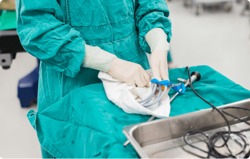
Thoracoscopy is a procedure usually used to check for issues with the pleura, a thin lining that covers the lungs. Also known as pleuroscopy, the procedure involves inserting a tube with a tiny camera through a small incision between the ribs. This allows doctors to view inside the chest. Unlike surgeries, thoracoscopy is minimally invasive. It helps with diagnosing certain lung conditions.
Key Facts About Thoracoscopy
- The thoracoscope is a thin, flexible tube with a camera. It’s inserted into the patient and usually requires only mild sedation.
- Thoracoscopy usually has a faster recovery and less pain than open surgery.
- It helps doctors diagnose, and in some cases, treat certain lung issues.
Thoracoscopy is a targeted approach. It’s often recommended when imaging tests like X-rays and CT scans don’t provide enough information. It can be especially useful when diseases such as mesothelioma are suspected. It’s a reliable way to detect and diagnose lung-related conditions.
The Role of Thoracoscopy in Diagnosing Mesothelioma
Thoracoscopy is widely considered the gold standard for diagnosing mesothelioma. Thoracoscopy is known for its high accuracy. A recent study in the European Respiratory Review shows it has a 91.5% accuracy rate for detecting fluid buildup in the chest from mesothelioma.
It can target the affected areas and provide detailed images to help determine the stage or extent of spread. It also allows doctors to take fluid or tissue samples for a biopsy. This particularly helps in diagnosing mesothelioma.
Dr. Jeffrey Velotta, thoracic surgeon at Kaiser Permanente, spoke with us about the value of thoracoscopy. He explains, “It’s important to have a thoracic surgeon or pulmonologist get in there and biopsy it to get tissue early on.”
Thoracoscopy for Mesothelioma Staging
A thoracoscopy can help doctors get a clearer picture of how far mesothelioma has progressed. A 2023 study in Cancers shows that collecting pleural fluid, during the procedure, helps find key biomarkers. These markers signify disease progression.
Staging mesothelioma helps doctors determine if the cancer is localized to an area, or if it has spread to other parts of the body. This aids in treatment planning, like surgery, radiation and chemo.
The more accurate the staging process, the higher the chances of improving the outcome. If tumors spread to nearby lymph nodes or other organs, doctors use this information as a guide to create a personalized treatment plan.
Get a Mesothelioma Treatment Guide

Find a Top Mesothelioma Doctor

Access Help Paying for Treatment

What to Expect With Thoracoscopy
First, you’ll meet with your surgeon to evaluate if thoracoscopy is suitable for you. At this meeting, your surgeon will explain the procedure. They’ll share details about prepping and recovery.
If you have pre-existing heart issues, you may need to have an additional meeting. A cardiologist may need to evaluate you before proceeding with the thoracoscopy.
Preparing for Your Thoracoscopy
It’s important to prepare for a thoracoscopy ahead of time to help ensure the procedure is successful. At the time of your consultation, your doctor will provide instructions on how to prepare. It can help to bring questions you may have to this meeting.
What To Ask Your Doctor
- When should I stop eating or drinking before my procedure?
- Should I stop taking any medications beforehand?
- Should I avoid using things like lotions or deodorant on my skin on the day of my procedure?
- Will I be able to manage my tasks independently after the procedure, or will I need assistance?
Your doctor will likely tell you to stop smoking at least 4 weeks before your thoracoscopy. Some hospitals will require a nicotine test before the procedure. If you’ve been smoking, your surgeon may cancel or reschedule your procedure.
Procedure
Before the procedure, your doctor may take a CT scan to identify where they need to investigate. This is important for your doctor to plan a thorough and targeted investigation.
Patients typically receive local anesthesia. This means you may stay awake and comfortable during your procedure. Once the anesthesia takes effect, you’ll be positioned to one side. A small incision will be made near your shoulder blade area. Your surgeon will insert an endoscope, a thin tube with a camera, through the incision. This will allow your doctor to get a clear view of the chest cavity.
If a biopsy is required, additional tools may be used to obtain samples. Once the procedure is complete, a temporary draining tube may be placed to drain any fluid or air buildup. The incision is then closed. The entire procedure generally takes around 45 to 90 minutes.

Connect with trusted specialists who truly care about your health. Get fast, stress-free appointment help.
Find a Doctor NowRecovering From a Thoracoscopy for Mesothelioma
Once your thoracoscopy is complete, you might be allowed to go home the same day. You may feel some soreness in the chest area, though. Sometimes patients need to stay overnight.
After Your Thoracoscopy
- Right after the procedure: You’ll be closely monitored, as the effects wear off quickly. There may be some pain in the chest area.
- Recovery at home: Get sufficient rest and avoid heavy lifting for a few days.
- Monitoring: Watch for any signs of infection, such as redness, swelling or fever. Contact your doctor right away if you experience these complications.
You’ll be provided detailed instructions to follow at home to help with the recovery process. Also, a follow-up visit will be scheduled. At your follow-up visit, your doctor will discuss the biopsy results with you and explain the next steps.
Risks and Complications of Thoracoscopy
Thoracoscopy is generally considered safe and effective. It has few risks and low complication rates. As a minimally invasive procedure, recovery time is usually shorter.
Studies show that there were major complications in fewer than 5% of cases. Minor complications were reported in just 1.5% of cases. In a 2022 retrospective, zero deaths occurred around the time of the procedure. This underscores its safety profile.
Potential Risks of Thoracoscopy
- Adverse reaction to anesthesia
- Air leak through the lung
- Pain or numbness
- Pneumonia
- Severe bleeding
- Wound infection
In another study in 2024, 141 patients were evaluated. There were zero cases of bleeding complications or persistent air leaks.
Complications are infrequent. However, patients with underlying conditions may be at higher risk. Discuss these risks with your surgeon.
Thoracoscopy vs. a VATS Procedure
Thoracoscopy and video-assisted thoracic surgery can both be used to diagnose mesothelioma. Though similar, the two procedures differ in important ways. Knowing these differences helps patients better prepare and can make informed decisions.
| Thoracoscopy | VATS | |
|---|---|---|
| Anesthesia | Local | General |
| Incisions | 1-2 small incisions | 3-4 small incisions |
| Purpose | Diagnostic | Diagnostic and surgical |
| Required instruments | Thoracoscope | Thoracoscope and additional instruments |
| Recovery setting | Outpatient, typically | Inpatient |
| Recovery time | Shorter, discharged same day | Longer, requires hospital stay |
Generally, VATS allows for more complex interventions. This can include surgical removal of tissue. This will necessitate a hospital admission. Both VATS and thoracoscopy allow for clear visualization and sample collection. VATS is preferred when more extensive procedures are needed.
Common Questions About Thoracoscopy for Mesothelioma
- Can I get a second opinion on next steps following my thoracoscopy results?
-
Absolutely! You can get a second opinion to confirm your thoracoscopy results and plan on next steps. The Patient Advocates at The Mesothelioma Center can match you with specialists who can offer a second opinion. They can also answer your questions about further care.
- What is the difference between a thoracoscopy and a thoracotomy?
-
Both thoracoscopy and thoracotomy allow doctors to view the chest cavity. The difference lies in the way the procedures are performed. Thoracoscopy is minimally invasive and involves small incisions. A thoracotomy is an open surgery with a larger incision for more direct access to the chest area. This is often used when more extensive treatment or tissue excision is required.
- How is a thoracoscopy different from a thoracentesis?
-
Both procedures involve the chest area, but serve vastly different purposes. A thoracentesis involves inserting a needle to drain excess fluid from the chest. It doesn’t provide visual access to the chest cavity like thoracoscopy does.
- Do patients with mesothelioma ever have more than one thoracoscopy procedure?
-
Yes, some patients with mesothelioma may have to undergo more than one thoracoscopy. Repeat procedures may be required to track disease progression, and evaluate responses to treatment.


